Szerző: admin | ápr 8, 2017 | Diffuse liver disease, Focal liver lesions
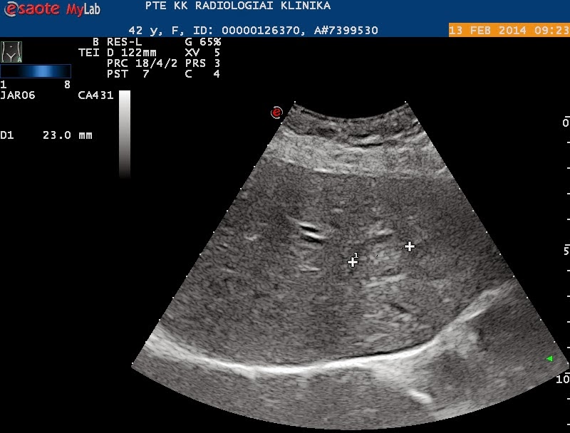
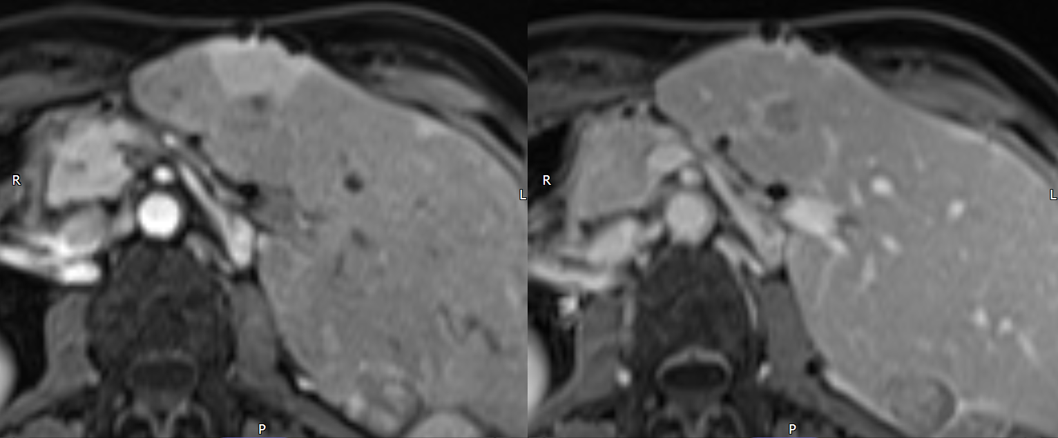
T1w C+ arterial and portal venousT1w and T2wRecurrent RCC with liver metastasis. The left liver lobe is disproportionately enlarged due to previous right lobectomy. Two intrahepatic mets are noted. The one in the midline is less obvious on the above images, but the...
Szerző: admin | szept 2, 2014 | Biliary tract, Focal liver lesions
35911
Szerző: admin | jún 15, 2012 | CEUS, Focal liver lesions
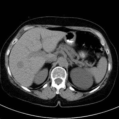
NECT: hypodens focal liver lesion in the right lobeCECT arterial phase: peripheral enhancementCECT venous phase: smaller lesion, brobable hemangiomaCECT late phase: no lesion seen (totally washed in?)CEUS ’29 (arterial): fast wash inCEUS over 2 min: total wash...

Szerző: admin | márc 20, 2012 | Focal liver lesions
CECT arterial phase; note the central feeding vesselthe lesion shows a bit unusual, slightly hypodense appearance in the venous phasecentral vessel and its branches in the coronal recon3D reconstruction shows the strong feeding artery of the FLL3D reconstruction...
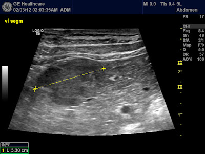
Szerző: admin | febr 4, 2012 | CEUS, Focal liver lesions, TIC
Perilesional hypervascularityTime-intensity-curves of to selected ROI (yellow – ring enhancement of the FLL, blue – normal liver parnchyma). Note the early enhancement of the metastases (time-to-peak).overlayed “reconstruction” of the...
